🇫🇷 Lire en Français | 🇪🇸Leer en Español | 🇧🇷Leia em português
This topic landed on my radar in an unexpected way. A colleague from Canada reached out to my former boss asking about canine maternal hydrops — a term that made me pause. When the article they shared mentioned a “serious lack of available information” about this condition in dogs, I had to dig deeper. What I found was startling: a single documented case report — published by Smith and Oaksford back in 1972 — was essentially all the veterinary literature had to offer.
That gap between what breeders experience in the field and what scientists have documented became the driving force behind this article. I’ve heard how quickly canine maternal hydrops can turn deadly when nobody recognizes it in time. My goal here is simple: give you the knowledge to spot it early, act fast, and potentially save your dog’s life — even if your vet has never heard of it.
- TL;DR: Key Takeaways
- What Is Canine Maternal Hydrops and How Does It Develop?
- What Should You Do If You Suspect Hydrops?
- What Tools and Supplies Should You Have Ready?
- What Warning Signs Should You Watch for During Pregnancy?
- You Have the Knowledge — Now Trust It
TL;DR: Key Takeaways
Canine maternal hydrops is a dangerous buildup of fluid in the fetal sacs during pregnancy that can kill both the mother and her puppies.
The two main types — hydrallantois (a placenta problem) and hydramnios (a fetal problem) — require different approaches to care.
A “rock-hard” belly, swollen vulva and hocks, blurry late-pregnancy X-rays, and extreme weight gain are the biggest red flags.
Ultrasound is the best diagnostic tool — specifically measuring the fluid depth around each puppy.
An emergency C-section is usually needed to save the mother’s life.
Your vet may not be familiar with this condition — you may need to bring the information to them, or ask them to consult a theriogenologist (reproductive specialist).
The condition is most common in first pregnancies and does not always repeat, but changing the stud for future breedings is strongly recommended.
What Is Canine Maternal Hydrops and How Does It Develop?
Hydrallantois vs. Hydramnios: Two Different Conditions
When breeders hear “hydrops,” they often think it’s one single condition. In reality, “dropsy of the fetal sacs” refers to two distinct problems. Understanding the difference matters because each one points to a different root cause — and that helps your vet choose the right approach.
Hydrallantois is the more common form. It happens when too much fluid builds up inside the allantoic sac — the outer fluid-filled membrane that surrounds each puppy. Think of the allantoic sac like a water balloon that wraps around the puppy’s inner protective bubble. When the placenta stops working properly, this outer balloon fills with way too much watery fluid.
Hydramnios is less common and involves the amniotic sac — the inner bubble closest to the puppy. This form is usually caused by a problem with the fetus itself, often a genetic or developmental defect that prevents the puppy from swallowing or processing amniotic fluid normally. In dogs, hydramnios is closely linked to fetal anasarca, sometimes called “water puppy syndrome.”
| Feature | Hydrallantois | Hydramnios |
|---|---|---|
| Which sac is affected? | Allantoic sac (outer membrane) | Amniotic sac (inner membrane) |
| Root cause | Placenta stops working properly | Fetal defect (genetic or developmental) |
| How common? | Most common form of hydrops | Less frequently reported in dogs |
| Type of fluid | Clear, watery, amber-colored | Thick, viscous, syrupy |
| Often associated with | Blood vessel failure in the placenta | Fetal anasarca (water puppy syndrome) |
How the Placenta Fails: Normal Fluid vs. Dangerous Hydrops
To understand canine maternal hydrops, it helps to know a little about how your dog’s placenta works. Dogs have what scientists call a zonary placenta — picture it like a wide belt or band that wraps around each puppy inside the uterus. This belt is lined with special cells called decidual cells that act as gatekeepers, controlling what passes between the mother’s blood and the puppy’s blood.
Here’s what makes the canine placenta unique: unlike in humans, it doesn’t produce its own hormones. These gatekeeper cells rely entirely on progesterone (the pregnancy hormone) produced by the ovaries to stay alive and functional. As long as progesterone levels stay healthy, the gatekeepers manage fluid exchange perfectly — just the right amount flows in and out.
In a healthy pregnancy, the fluid surrounding each puppy follows a predictable pattern. It gradually increases, reaching its peak around week six of gestation, and then naturally decreases toward delivery. Your vet can measure this fluid depth on ultrasound — it’s called the allantoamniotic fluid depth. When that measurement keeps rising after week six instead of falling, that’s a clear warning sign.
When hydrallantois strikes, those gatekeeper cells become compromised. The blood vessels in the placenta lose their ability to regulate what passes through — they become too permeable, like a garden hose full of tiny holes. Instead of controlled fluid exchange, blood plasma floods into the fetal sacs uncontrollably. The resulting fluid is not normal amniotic fluid — it’s a clear, watery, amber-colored liquid that is essentially filtered blood.
The volume can become so massive that it physically crushes the mother’s internal organs — pressing against her diaphragm so she can’t breathe, squeezing her stomach so she can’t eat, and compressing major blood vessels. This is what makes canine maternal hydrops so dangerous: it can lead to breathing failure, severe swelling throughout the body, and even fatal heart failure.
This is why asking your vet to specifically measure fluid depth on ultrasound — not just do a quick pregnancy check — is so important. A rising fluid measurement after week six is one of the earliest detectable signs that the placenta is failing.
| Measurement | Normal Pregnancy | Hydrops Pregnancy |
|---|---|---|
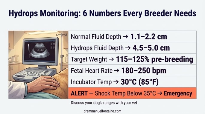
| Fluid depth per puppy | 1.1 to 2.2 cm (0.4 to 0.9 in) | 4.5 to 5.0 cm (1.8 to 2.0 in) or more |
| Fluid trend after week 6 | Gradually decreases toward delivery | Continues to increase — the key red flag |
| Fluid appearance | Normal amniotic fluid | Clear, watery, amber-colored (filtered blood plasma) |
| Abdominal feel | Soft with some give | Rock-hard, extremely tense |
| X-ray clarity (day 55–56) | Clear fetal skeletons visible | Blurry image because fluid scatters the beam |
What Cattle Research Teaches Us About Canine Hydrops
Here’s a fascinating detail from veterinary research: much of what scientists understand about hydrops actually comes from cattle medicine. Because this condition is so rare and poorly documented in dogs, the bovine world — where hydrops is extensively studied — provides critical scientific insight.
Cattle have a different placental structure than dogs, but bovine hydrallantois follows the same basic pattern: the attachment sites between mother and fetus (called caruncles in cows) fail to form properly. The placenta tries to compensate by growing larger, but this enlarged placenta is dysfunctional and leaks fluid uncontrollably.
The most eye-opening finding comes from cloned cattle. In cows produced through cloning, hydrallantois occurs at rates between 25% and 75%. That’s an enormous number. What this tells scientists is that hydrops is fundamentally a disorder of “placental programming.” Errors during the earliest stages of embryonic development — particularly in how genes are switched on and off (a process called epigenetics, which you can think of as the “instruction manual” for which genes to use and which to ignore) — lead to catastrophic problems in how the placenta builds its blood vessels and manages fluid.
For dog breeders, the takeaway is powerful: canine maternal hydrops likely starts long before you ever see symptoms. It begins at the very foundation of pregnancy, when the embryo’s placenta is first being built. Ask your vet about whether early ultrasound monitoring at regular intervals could help detect fluid changes before they become an emergency.
| Insight from Cattle | What It Means for Dog Breeders |
|---|---|
| Hydrops is linked to placental attachment failure | The problem starts early in pregnancy, not late |
| Cloning causes hydrops in 25–75% of cattle pregnancies | Errors in gene activation during early development are a major driver |
| Compensatory placental growth makes it worse | A bigger placenta does not mean a healthier pregnancy |
| Bovine research provides treatment frameworks | Protocols from cattle medicine can guide how we manage canine cases |
What Should You Do If You Suspect Hydrops?
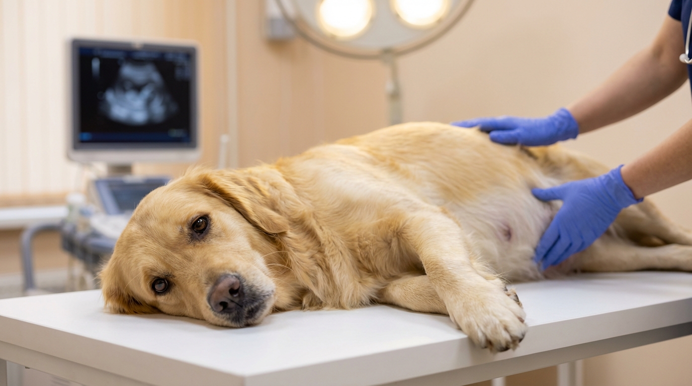
Monitor Your Pregnant Dog Like a Detective
The single most important thing you can do is pay close attention to your pregnant dog’s body throughout gestation. Canine maternal hydrops doesn’t announce itself with a single dramatic sign. It creeps in gradually, and the breeders who catch it early are the ones who were watching closely from the start.
Start by knowing your baseline. Get an early ultrasound to estimate your puppy count. From there, track your dog’s weight regularly.
By the end of gestation, a pregnant dog should typically weigh 115 to 125 percent of her optimal pre-breeding body weight.
Weight gain does not occur uniformly throughout pregnancy. In most bitches, the increase becomes more noticeable and progresses in a relatively linear manner from approximately day 40 of gestation until whelping.
This matters because body weight is not just a number on a scale. It is a practical monitoring tool. If you know her ideal starting weight, you can track her weekly gain and verify that gestation is progressing within expected physiological limits. Too little gain may signal inadequate nutritional intake or small litter size. Excessive gain may reflect overfeeding or metabolic imbalance.
In other words, weight monitoring during gestation is not optional. It is a clinical indicator.
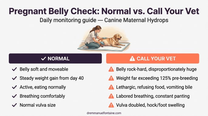
Feel her belly every day. A normal pregnant abdomen has some softness — you can gently press and feel movement. A hydrops belly becomes rock-hard and disproportionately huge, with absolutely no room left for growth. Also watch for swelling in her vulva (which may double in size), her hocks, and her feet. And pay attention to her behavior: lethargy, refusing food, vomiting bile, and heavy panting are all alarm bells.
| What to Monitor | Normal | Red Flag |
|---|---|---|
| Weight gain | Proportionate to puppy count (about 1 kg / 2 lbs per puppy) | Weight gain far exceeding expected amount |
| Abdominal feel | Soft with gentle give when pressed | Rock-hard, tense, no room for growth |
| Vulva size | Mild swelling in late pregnancy | Doubled in size, severe swelling |
| Breathing | Normal, occasional panting | Labored breathing, constant panting |
| Appetite and energy | Eating well, active | Refusing food, vomiting bile, lethargy |
| Vaginal discharge | None until close to labor | Clear watery fluid leaking early |
Get the Right Diagnostics at the Vet
When you walk into your vet’s office suspecting hydrops, you need to know exactly what to ask for. Remember, many veterinarians have never encountered this condition. Being prepared with specific requests can make the difference between a timely diagnosis and dangerous delays.
The gold standard diagnostic tool is a transabdominal ultrasound. But not just any quick scan — you need your vet to specifically look for abnormal fluid accumulation between the fetal membranes and measure the fluid depth around each puppy. As we covered in the WHAT TO KNOW section, normal fluid depth runs between 1.1 and 2.2 cm (0.4 to 0.9 in). Hydrops-affected puppies in documented cases showed depths of 4.5 to 5 cm (1.8 to 2.0 in) — more than double normal.
While the ultrasound is running, ask the vet to check fetal heart rates. Healthy puppy hearts beat between 180 and 250 beats per minute. If heart rates drop significantly below that range (a condition called bradycardia, meaning “slow heartbeat”), the puppies are in serious distress. And if you’re doing late-gestation X-rays around day 55–56 to count skulls, watch for a “blurry” film. That haze happens because the massive volume of fluid scatters the X-ray beam, hiding the fetal skeletons.
| Diagnostic Tool | What to Request | What It Reveals |
|---|---|---|
| Transabdominal ultrasound | Measure fluid depth around each puppy (should be 1.1–2.2 cm / 0.4–0.9 in normally) | Depths above 4.5 cm (1.8 in) suggest hydrops |
| Fetal heart rate check | Monitor beats per minute for each puppy | Below 180 bpm = serious fetal distress |
| Late-gestation X-rays | X-rays around day 55–56 for skull count | Blurry image = fluid scattering the beam |
Choose the Right Treatment Path
Once canine maternal hydrops is confirmed, you and your vet face a critical decision. The right path depends on how far along the pregnancy is, how stable the mother is, and how the puppies are doing.
In almost all cases, a C-section is ultimately required. The mother should not go through the strain of natural labor with hydrops. If fetal heart rates drop or the mother starts crashing, this becomes an emergency procedure that requires special consideration. Indeed, when the surgeon opens the uterus, the sudden loss of massive fluid volumes can send the mother into fatal shock (called hypovolemic shock, meaning her body loses so much fluid that her heart can’t pump enough blood) within minutes.

If severe hydrops is diagnosed very early and the mother’s life is in immediate danger, pregnancy termination may be the safest option to save the dam.
What Tools and Supplies Should You Have Ready?
Home Monitoring Equipment
Being prepared for canine maternal hydrops starts with having the right monitoring tools before your female is even bred. These are items every serious breeding program should already own, but they become absolutely essential when hydrops is a possibility.
An accurate digital scale is your first line of defense. As we discussed earlier, tracking your dog’s weight against the expected norm for her litter size is the earliest way to catch disproportionate gain. Without a scale, you’re guessing — and with hydrops, guessing can be fatal.
A digital rectal thermometer helps you track the pre-labor temperature drop. When your dog’s temperature falls from its normal 38 °C (101 °F) down below 37 °C (99 °F), labor should begin within about 24 hours. This timing becomes critical if you’re managing a hydrops pregnancy and trying to coordinate an emergency C-section with your vet.
Build Your Veterinary Support Team
This is where canine maternal hydrops gets tricky, and it’s something I emphasize with every breeder I consult: your regular vet may not be familiar with this condition.
And it’s a reflection of how poorly this condition has been documented in veterinary literature. As I mentioned in the introduction, there was essentially one published case report for decades. You need a veterinary team that is willing to listen when you raise concerns, even about conditions they haven’t personally seen. Better yet, establish a relationship with a board-certified theriogenologist (a veterinary reproductive specialist) who can consult by phone or telemedicine if your local clinic needs guidance.
Have your emergency plan ready before you need it.
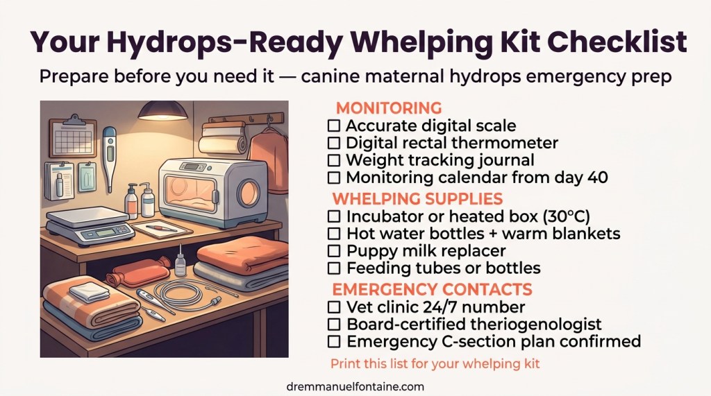
Stock Your Emergency Whelping Kit
Puppies born from hydrops pregnancies are often highly compromised. Many are premature, some may have concurrent water puppy syndrome (anasarca), and neonatal mortality is exceptionally high. When I advise breeders preparing for a possible hydrops delivery, I tell them to prepare for the hardest whelping of their life.
Your whelping area needs an incubator or heated box set to exactly 30 °C (85 °F), stocked with hot water bottles and a constant supply of warm blankets. These puppies are extremely vulnerable to fatal chilling.
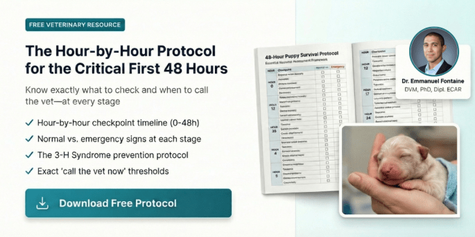
Before whelping, take the time to review your neonatal resuscitation protocol.
This is not something you improvise at 2 a.m. when a non-breathing puppy is in your hands. Resuscitation is procedural. It requires clarity, speed, and the right sequence of actions.
Knowing exactly what to do, in what order, and at which thresholds can be the difference between panic and precision.
Preparation is part of perinatal management. Not an afterthought.
Stock puppy milk replacer and feeding tubes or bottles because the dam’s milk may dry up, fail to come in, or she may be too exhausted to nurse. You should be prepared to tube-feed or bottle-feed the entire litter around the clock.
What Warning Signs Should You Watch for During Pregnancy?
Early Warning Signs You Should Never Ignore
Catching canine maternal hydrops early dramatically improves outcomes for both the mother and her puppies. The challenge is that the early signs can easily be mistaken for a normal large litter. Because dogs naturally carry multiple puppies, extreme abdominal size doesn’t automatically raise alarms — and that’s exactly why this condition slips past so many breeders and vets.
You already have the tools to catch this early. Remember the weight tracking and belly checks? Those daily monitoring habits are your first line of defense here. When your dog’s weight is climbing far beyond the expected range, and her abdomen feels rock-hard instead of soft and moveable, you have strong reason to be concerned.
On the diagnostic side, remember what we covered about ultrasound measurements: fluid depths significantly above the normal 1.1–2.2 cm (0.4–0.9 in) range — especially readings of 4.5 cm (1.8 in) or higher — are direct evidence of hydrops. And if late-gestation X-rays come back looking blurry instead of showing crisp fetal skeletons, that’s another diagnostic red flag pointing directly to excessive fluid.
| Early Sign | What It Looks Like | Why It Matters |
|---|---|---|
| Disproportionate weight gain | Weight far exceeding breed norm for litter size | Earliest measurable warning (use your scale and puppy count) |
| Rock-hard belly | Abdomen feels extremely tense with no give | Indicates dangerous fluid pressure, not just a large litter |
| Abnormal ultrasound fluid depth | Readings of 4.5–5+ cm (1.8–2.0+ in) per puppy (normal: 1.1–2.2 cm / 0.4–0.9 in) | Directly measures the excess fluid causing the condition |
| Blurry late-gestation X-rays | Fetal skeletons hard to see on day 55–56 films | Massive fluid volume scatters the X-ray beam |
Emergency Danger Signs That Need Immediate Action
Some signs tell you that your dog’s body is running out of time to compensate. When you see any of these, you need to be at the veterinary clinic immediately — not tomorrow morning, not after the weekend. Right now.
Severe peripheral edema (sudden dramatic swelling) in the limbs, hocks, feet, and especially the vulva means fluid is overwhelming the entire body, not just the uterus. Labored breathing and constant panting signal that the massive fluid volume has compressed the diaphragm — exactly the organ compression we explained earlier — and the mother is struggling to get enough oxygen.
Watch for systemic decline: extreme lethargy, complete loss of appetite, depression, and heavy vomiting of bile as the delivery date approaches. If you see clear watery fluid leaking from the vulva before labor should start, the fetal membranes may be failing. And the most alarming indicator of all — pale gums, dehydration, and a body temperature dropping to 35 °C (95 °F) — means the mother is going into shock. This is the moment when the aggressive IV fluid protocol from our treatment discussion becomes a matter of life and death.
| Danger Sign | What It Means | Action Required |
|---|---|---|
| Severe limb/vulva swelling | Fluid overwhelming the entire body | Call emergency vet immediately |
| Labored breathing, constant panting | Diaphragm compressed by fluid buildup | Prepare for emergency C-section |
| Vomiting bile, complete loss of appetite | Internal organs compressed, body failing | Urgent veterinary evaluation |
| Clear fluid leaking from vulva | Fetal membranes failing prematurely | Emergency vet visit — do not wait |
| Pale gums, body temperature at 35 °C (95 °F) | Mother going into shock | Life-threatening emergency — surgery now |
| Fetal heart rate below 180 bpm | Puppies in severe distress | Emergency C-section required |
Signs Treatment Is Working or Failing
After the C-section, monitoring doesn’t stop — it actually intensifies. Knowing the difference between recovery and failure helps you and your vet make fast decisions in the critical hours and days that follow surgery.
The primary sign that treatment is working is maternal stability after surgery. The mother’s vital signs should stabilize. Her color should return, her breathing should ease, and she should gradually regain energy.
For the puppies, success looks like neonates responding to active airway clearance, warming up in the 30 °C (85 °F) incubator from your whelping kit, and successfully latching on to nurse or accepting milk replacer. But be prepared: even with a successful maternal outcome, neonatal mortality remains very high with hydrops litters.
Watch carefully for post-surgical eclampsia (milk fever) in the mother. If her milk comes in suddenly after surgery, calcium levels can crash fast. Muscle weakness, tremors, temperature spikes, and behavioral changes are warning signs that require immediate calcium supplementation — this is exactly why we included calcium in the emergency whelping kit. And for future breeding decisions, note that canine maternal hydrops is most common in first pregnancies and does not always recur. However, because genetic factors and specific sire-dam combinations may play a role, changing the stud for future breedings is strongly recommended.
| Indicator | Treatment Working | Treatment Failing |
|---|---|---|
| Mother’s vital signs after surgery | Stable color, normal breathing, gradual energy return | Pale gums, continued labored breathing, shock symptoms |
| Newborn puppy response | Puppies respond to warming, latch to nurse | Puppies dying shortly after birth, failing to thrive |
| Mother’s calcium levels | Normal nursing without complications | Muscle tremors, temperature spikes, behavioral changes (eclampsia / milk fever) |
| Future breeding outlook | Condition may not recur in next pregnancy | Same sire-dam combination produces hydrops again |
You Have the Knowledge — Now Trust It
Canine maternal hydrops is rare, poorly documented, and frightening when it happens. But you just learned something that puts you ahead of the vast majority of breeders — and ahead of many veterinarians, too. You now understand what this condition is, how the placenta fails, how to recognize the early warning signs, what diagnostics to request, how to prepare your whelping kit, and what emergency signs demand immediate action.
That knowledge is powerful. It transforms you from someone who might have dismissed a rock-hard belly as “just a big litter” into someone who can potentially save their dog’s life. It turns you into a medical advocate for your animals — someone who walks into the vet’s office with a monitoring journal, specific ultrasound requests, and a C-section advocacy checklist ready to go.
Remember: the biggest danger with canine maternal hydrops isn’t the condition itself — it’s not knowing it exists. You no longer have that problem. Share this knowledge with the breeders in your community. Talk to your vet about it. Keep monitoring, keep learning, and trust your instincts when something doesn’t look right. Your dogs are counting on you, and now you’re ready.

I just had a hydrops litter born yesterday. 9 puppies delivered by C-section. Heart rates extremely low on ultrasound. Akita bitch had excessively enlarged vulva and had been leaking for 1 week. She hadn’t eaten in 5 days but was drinking. Vomiting bile the last 24 hours. 61 days post first breeding. Temp drop from 101 to 99 followed by non-productive nesting behavior for 8 hours. Normally 78 lb bitch weighed in at 122 lbs. Uterus had huge volumes of fluid. When the first horn was pulled out, it was distended at least twice normal size and you could see movement of puppies sloshing around in it (which was also seen in ultrasound). When uterine wall was opened, it was like someone dumped a 1 gallon bucket for both horns. Puppies were extremely slow to thrive, but okay now. Bitch’s mammary development dried up post surgery and was given Domperidone and was back in 5 hours later but she spiked 103 and showed all the behavior changes of dropped calcium levels with the sudden milk production, but she is responding to high volumes of oral calcium.
This is the second I have had … first was a litter of one puppy about 8 years ago.
I am happy to put you in touch with my veterinarian.
LikeLike