Ten days ago I was lecturing at the vet colleges of Saskatoon and Calgary. The talk I had prepared was named: “Clinical cases in canine and feline obstetrics: should I be afraid when a breeder called in the middle of the night?”. Wonder what our goal was here? I just wanted to show the vet students that, when they will be in practice, they should not be afraid of dealing with small animal obstetrics. We now have the veterinary tools to properly answer questions related to canine and feline parturition. And that’s good because when they will be in practice, this is something they will definitely see !
These talks are always a good opportunity to discuss with vet students. During this trip out West then, in both schools I was asked the same question: “During whelping, is it possible to assess the presentation of the puppy/kitten under ultrasounds?”.
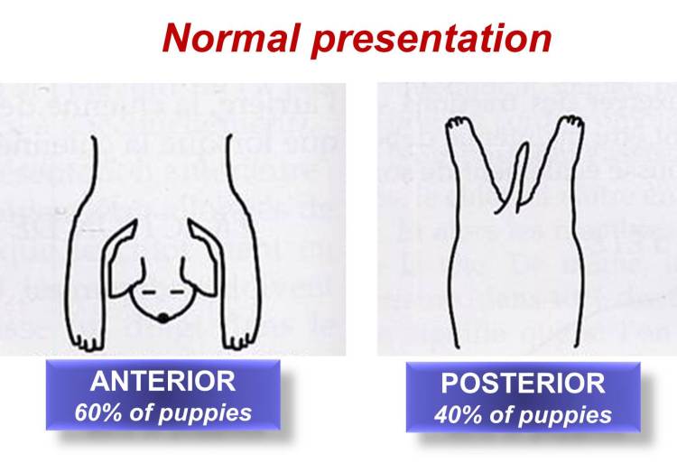
Let’s start with some definition: in obstetrics, presentation signifies the relationship between the longitudinal axis of the fetus and the maternal birth canal. Normal presentation is usually longitudinal (see picture below). What we don’t want to deal with? Tranverse presentation, where in fact the spine of a puppy/kitten becomes perpendicular with the birth canal.
When a bitch or a queen comes to the clinic for dystocia (=difficulty to give birth), this is always something to assess. Why? Look at the X-ray below, when we see this picture during parturition, we know that there is no way the mother will be able to expulse naturally the puppy/kitten. A transverse presentation is definitely an indication for C-section.

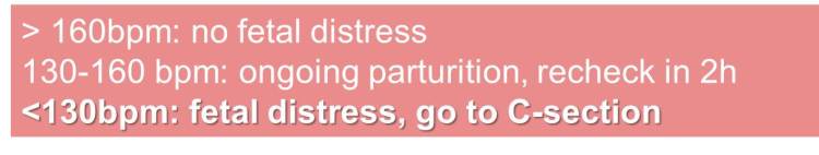
Presentation assessment is performed through X-rays, since this technique will give us a complete view of the abdominal cavity. Why do we speak about ultrasounds then? I always use ultrasounds in fact when dealing with an obstetrical case, this is in fact usually the first thing I would do after examining the bitch. It helps us determining the heart beat frequency of the puppies/kittens, which is also an important element when it comes to take a decision oh how to properly handle the case (see Table below). However, ultrasounds allow us to visualize only sections of the abdomen, and will never give us the “big picture” in terms of presentation. That’s why they will never replace X-rays, and both should be part of the veterinary approach in small animal obstetrics.